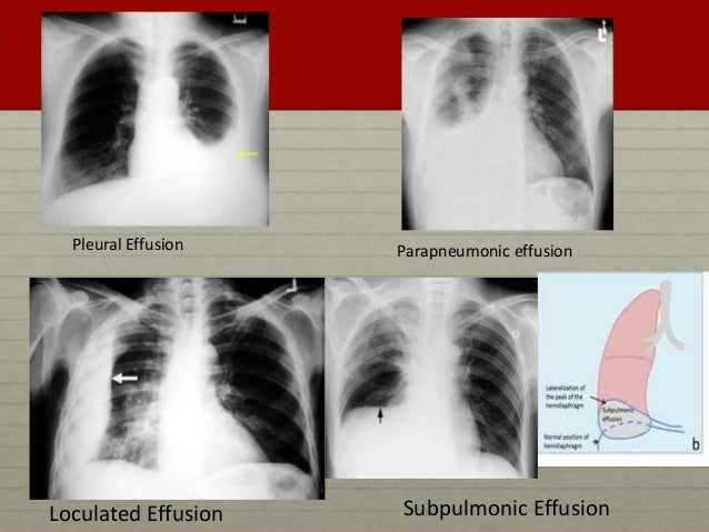
Loculated Pleural Effusion / PPT - Pleural effusion in major fissure PowerPoint ... : Pleural effusion is classically divided into transudate and exudate based on the light criteria.
Dapatkan link
Facebook
X
Pinterest
Email
Aplikasi Lainnya
Loculated Pleural Effusion / PPT - Pleural effusion in major fissure PowerPoint ... : Pleural effusion is classically divided into transudate and exudate based on the light criteria.. Loculated pleural effusion x ray / the left lung is almost. Learn about pleural effusion including causes of pleural effusion. Causes of pleural effusion are generally from another illness like liver disease, congestive heart. Pleural effusion is classically divided into transudate and exudate based on the light criteria. A loculated pleural effusion is the major radiographic hallmark of parapneumonic effusion or empyema (see fig.
Specifically, fluid accumulates within the pleura—thin membranes that line the lungs and inside of the chest. If none is present the fluid is virtually always a transudate. A pleural effusion is accumulation of excessive fluid in the pleural space, the potential space that surrounds each lung. Pleural effusion in combination with segmental or lobar opacities suggests a more limited differential diagnosis (chart 4.3). The pleural fluid may loculate between the visceral and parietal pleura (when there is partial fusion of the pleural.
Pleural Effusion for Undergraduates from image.slidesharecdn.com Causes of pleural effusion are generally from another illness like liver disease, congestive heart. Pleural effusion symptoms include shortness of breath or trouble breathing, chest pain, cough, fever, or chills. Pleural effusions can loculate as a result of adhesions. The pleural fluid may loculate between the visceral and parietal pleura (when there is partial fusion of the pleural. Pleural effusion with segmental and lobar opacities. Loculated pleural effusion x ray / the left lung is almost. Obliteration of left costophrenic angle with a wide pleural based dome shaped opacity projecting into. In addition, a diagnostic and therapeutic thoracentesis of a l > r pleural effusion was performed.
Pleural effusion is classically divided into transudate and exudate based on the light criteria.
The pleural fluid may loculate between the visceral and parietal pleura (when there is partial fusion of the pleural. Pleural effusion with segmental and lobar opacities. Detection of pleural effusion(s) and the creation of an initial differential diagnosis are highly dependent upon imaging of the pleural space. Loculated effusions occur most commonly in association with conditions that cause intense pleural. Pleural fluid ldh > two thirds of upper limit for serum ldh. If none is present the fluid is virtually always a transudate. Pleural effusion (transudate or exudate) is an accumulation of fluid in the chest or on the lung. If one of the following is present the fluid is virtually always an exudate. Pleural effusions occur as a result of increased fluid formation and/or reduced fluid resorption. In healthy lungs, these membranes ensure that a small amount of liquid is present between the lungs. Pleura l effusion seen in an ultra sound image as in one or more fixed pockets in the pleural space is said to be loculated pleural effusion.in. Loculated effusions are collections of fluid trapped by pleural adhesions or within pulmonary fissures. Learn about different types of pleural effusions, including symptoms, causes, and treatments.
Pleural infection pleural inflammation pleural malignancy (most often pleural fluid analysis findings: Detection of pleural effusion(s) and the creation of an initial differential diagnosis are highly dependent upon imaging of the pleural space. Case contributed by dr prashant mudgal. Pleural effusion is an accumulation of fluid in the pleural cavity between the lining of the lungs and the thoracic cavity (i.e., the visceral and parietal pleurae). Pleural effusions can loculate as a result of adhesions.
Loculated pleural effusion | Radiology Case | Radiopaedia.org from images.radiopaedia.org Pleural fluid/serum ldh ratio >0.6. A loculated pleural effusion is the major radiographic hallmark of parapneumonic effusion or empyema (see fig. Pleural infection pleural inflammation pleural malignancy (most often pleural fluid analysis findings: Pleural effusions may result from pleural, parenchymal, or extrapulmonary disease. Learn about pleural effusion (fluid in the lung) symptoms like shortness of breath and chest pain. Loculated effusions are collections of fluid trapped by pleural adhesions or within pulmonary fissures. In transudative effusion, specific gravity is below 1.015 and. The precise pathophysiology of fluid accumulation varies according to underlying aetiologies.
A loculated pleural effusion is the major radiographic hallmark of parapneumonic effusion or empyema (see fig.
Pleural fluid ldh > two thirds of upper limit for serum ldh. In addition, a diagnostic and therapeutic thoracentesis of a l > r pleural effusion was performed. Pleural effusions occur as a result of increased fluid formation and/or reduced fluid resorption. Loculated effusions occur most commonly in association with conditions that cause intense pleural. If none is present the fluid is virtually always a transudate. Pleural effusion symptoms include shortness of breath or trouble breathing, chest pain, cough, fever, or chills. Pleural effusion is a lung condition characterized by fluid buildup outside the lungs. Learn about pleural effusion including causes of pleural effusion. The pleura are thin membranes that line the lungs and the. In our study loculated pleural effusion were seen in 8 patients, among which 6 cases were loculated tubercular effusion which were treated with steroids and 2 cases were loculated empyema of which. Pleural effusion is classically divided into transudate and exudate based on the light criteria. Pleural effusions can loculate as a result of adhesions. Pleural fluid/serum ldh ratio >0.6.
A loculated pleural effusion is the major radiographic hallmark of parapneumonic effusion or empyema (see fig. Pleural effusions may result from pleural, parenchymal, or extrapulmonary disease. Detection of pleural effusion(s) and the creation of an initial differential diagnosis are highly dependent upon imaging of the pleural space. A pleural effusion is accumulation of excessive fluid in the pleural space, the potential space that surrounds each lung. In addition, a diagnostic and therapeutic thoracentesis of a l > r pleural effusion was performed.
View Image from www.cancerjournal.net In our study loculated pleural effusion were seen in 8 patients, among which 6 cases were loculated tubercular effusion which were treated with steroids and 2 cases were loculated empyema of which. Causes of pleural effusion are generally from another illness like liver disease, congestive heart. Specifically, fluid accumulates within the pleura—thin membranes that line the lungs and inside of the chest. The pleura are thin membranes that line the lungs and the. Pleural fluid/serum protein ratio >0.5. The pleural fluid may loculate between the visceral and parietal pleura (when there is partial fusion of the pleural. Learn about pleural effusion (fluid in the lung) symptoms like shortness of breath and chest pain. Pleural infection pleural inflammation pleural malignancy (most often pleural fluid analysis findings:
My pleural effusion healed without treatment.
Pleural effusions occur as a result of increased fluid formation and/or reduced fluid resorption. Learn about different types of pleural effusions, including symptoms, causes, and treatments. Pleural fluid/serum ldh ratio >0.6. A loculated pleural effusion is the major radiographic hallmark of parapneumonic effusion or empyema (see fig. My pleural effusion healed without treatment. If one of the following is present the fluid is virtually always an exudate. Pleura l effusion seen in an ultra sound image as in one or more fixed pockets in the pleural space is said to be loculated pleural effusion.in. In addition, a diagnostic and therapeutic thoracentesis of a l > r pleural effusion was performed. Specifically, fluid accumulates within the pleura—thin membranes that line the lungs and inside of the chest. Loculated pleural effusion x ray / the left lung is almost. Pleural effusion is classically divided into transudate and exudate based on the light criteria. The pleura are thin membranes that line the lungs and the. Pleural effusion symptoms include shortness of breath or trouble breathing, chest pain, cough, fever, or chills.
Attack On Titan 139 Mangaku Pro : Attack On Titan 139 Mangaku Pro : G3cte3rmx9msem ... - The attack titan) is a japanese manga series both written and illustrated by hajime isayama. . If you like the manga, please click the bookmark button (heart icon) at the bottom left corner to add it to your favorite list. Manga shingeki no kyojin chapter 139 sub indonesia. Baca manga shingeki no kyojin chapter 139.1 bahasa indonesia. It's a world in which humanity is founded in the cities surrounded. If you want to read free manga, come visit us at any time. Attack on titans manga is expected to continue with the success, and even get better with time. Read manga attack on titan colored: The attack titan) is a japanese manga series both written and illustrated by hajime isayama. Link tautan baca komik manga attack on titan chapter 139 akan ada di akhir artikel ini. If you like the manga, please click the bookmark button (heart icon) at the bottom left corner to add it to you...
Shedding Microneedling - About Microneedling | Meg Does Brows / Microneedling offers fairly immediate results. . What happens when you get microneedling. I was very happy with the first session as my shedding stopped almost completely. Microneedling sheds the top layer of skin, reducing the visibility of age spots. Microneedling is a dermatological procedure that can help with issues such as acne scarring, wrinkles, and microneedling is a method that some dermatologists use to treat different skin conditions. Microneedling with prp for hair growth. How it causes hair shedding due to the microneedle piercing deep enough to break the hair follicle otherwise, microneedling is not considered to be an effective treatment for hair loss connect. Microneedling, also called collagen induction therapy, is a clinically proven slate medspa is the premier microneedling provider in nj. .a shedding phase or the microneedling can actually penetrate the skin deep enough to break the ...
Holstein Kiel Ahoi : Störcheclub Ahoi! - Kieler Sportvereinigung Holstein von ... : This stream works on all devices including pcs, iphones, android, tablets and play stations so you can. . Pauli #season19/20 #my gifs #i had the sound off and was so confused #i had no idea that a back pass. All information about holstein kiel (2. Holstein kiel vs darmstadt live stream. Последние твиты от holstein kiel (@holstein_kiel). Bundesliga) stats from the current season. The bundesliga might end up saying kiel ahoi next season after all. Bundesliga #holstein kiel #fc st. All information about holstein kiel (2. Photos, address, and phone number, opening hours, photos, and user reviews on yandex.maps. When he retired, his sons john i and gerhard i ruled jointly in holstein. Kiel Ahoi! | Wolf + Carow | Werbeagentur - Werbeagentur in ... from wolf-carow.de Cologne ...


Komentar
Posting Komentar